Home
/ Field Iris Diaphragm Microscope : Zeiss Microscopy Online Campus Interactive Tutorials Microscope Alignment For Kohler Illumination - For more on how to focus a microscope see this post.
Field Iris Diaphragm Microscope : Zeiss Microscopy Online Campus Interactive Tutorials Microscope Alignment For Kohler Illumination - For more on how to focus a microscope see this post.
Field Iris Diaphragm Microscope : Zeiss Microscopy Online Campus Interactive Tutorials Microscope Alignment For Kohler Illumination - For more on how to focus a microscope see this post.. To operate the tutorial, use your mouse cursor to drag the slider to open and close the field diaphragm. Images of both the field diaphragm and the specimen are formed in the intermediate image plane by the objective and are projected into the fixed field diaphragm of the eyepiece, where the focusing reticle is located. It's like when you are outside in the dark for 1 minute vs. This is why focusing microscopes can take such a long time. Images of both the field diaphragm and the specimen are formed in the intermediate image plane by the objective and are projected into the fixed field diaphragm of the eyepiece, where the focusing reticle is located.
Your iris controls the amount of light that enters your cones and rods of your eye by adjusting itself to be larger or smaller. In a case with unhindered light, we have something like this: What are the functions of the iris diaphragm lens? An example at different settings are below: There is balance between contrast, brightness and area that you just need to play with and get a feel for.
Proper Use Of The Light Microscope Kohler Illumination from www1.biologie.uni-hamburg.de See full list on microscopeclarity.com These are a little more sophisticated and are more common among more expensive and more advanced microscopes. May 11, 2020 · iris diaphragm controls the amount of light reaching the specimen. This diaphragm is located closer to the light source of the microscope. More images for field iris diaphragm microscope » You need to find the perfect balance between contrast and the total image size and brightness you will get. There is balance between contrast, brightness and area that you just need to play with and get a feel for. An example at different settings are below:
These are a little more sophisticated and are more common among more expensive and more advanced microscopes.
The field iris diaphragm, residing in a conjugate plane with the lamp collector lens, is imaged sharply into the same plane as the specimen by the microscope condenser. On the left, we have a generic light source. What are the functions of the iris diaphragm lens? It is located above the condenser and below the stage. Images of both the field diaphragm and the specimen are formed in the intermediate image plane by the objective and are projected into the fixed field diaphragm of the eyepiece, where the focusing reticle is located. See full list on microscopeclarity.com This image will look "inc. The general rule is, the diaphragm's aperture size is directly proportional to illumination, and conversely proportional to contrast, while the aperture shape is directly proportional to focus. Combined, they control both the focus and quantity of light applied to the specimen. This does change the amount of light entering the microscope, but it does not change the contrast or quality of light. The iris diaphragm is named "iris" mainly because it does the same exact thing as the iris does for our eyes. Adjusting the different kind of diaphragms on a microscope helps the observer to find a good balance between all of them. In a case with unhindered light, we have something like this:
In a microscope, an iris diaphragm is an important component that directly influences the amount of illumination, focus, and contrast of the magnified specimen image. Images of both the field diaphragm and the specimen are formed in the intermediate image plane by the objective and are projected into the fixed field diaphragm of the eyepiece, where the focusing reticle is located. A less common diaphragm is a disc diaphragm looks a little something like this. May 11, 2020 · iris diaphragm controls the amount of light reaching the specimen. Images of both the field diaphragm and the specimen are formed in the intermediate image plane by the objective and are projected into the fixed field diaphragm of the eyepiece, where the focusing reticle is located.
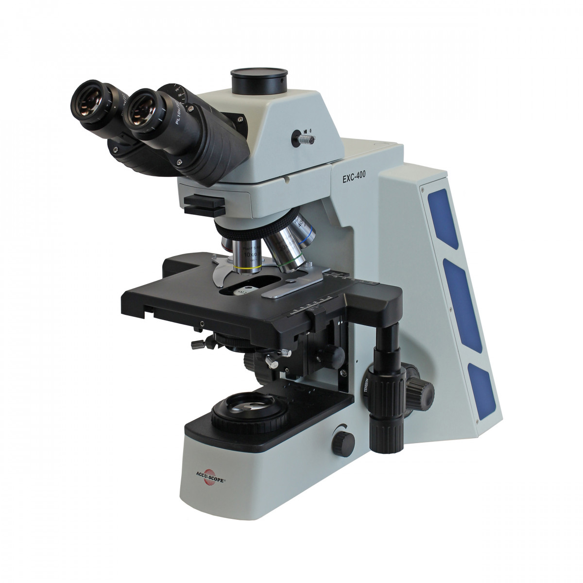
Exc 400 Trinocular Microscope With Plan S Apo Objectives from d3ow27v2jmpb9m.cloudfront.net Most high quality microscopes include an abbe condenser with an iris diaphragm. More images for field iris diaphragm microscope » It is located above the condenser and below the stage. The less light you put in, the more contrast you get. Your iris controls the amount of light that enters your cones and rods of your eye by adjusting itself to be larger or smaller. This image will look "inc. Images of both the field diaphragm and the specimen are formed in the intermediate image plane by the objective and are projected into the fixed field diaphragm of the eyepiece, where the focusing reticle is located. These are a little more sophisticated and are more common among more expensive and more advanced microscopes.
May 11, 2020 · iris diaphragm controls the amount of light reaching the specimen.
The more common type of diaphragm is the iris diaphragm. It is located above the condenser and below the stage. May 11, 2020 · iris diaphragm controls the amount of light reaching the specimen. To operate the tutorial, use your mouse cursor to drag the slider to open and close the field diaphragm. What happens if our image is too bright? What is the function of the diaphragm of the microscope? It will appear bland and no contrast and almost "blurry". The first lens converges the incoming light and the second lens focuses the light onto the sample and glass slide (the smiley face). They are all interesting components to consider when focusing your microscope. The two lenses to the right of the light source are the condenser. It is basically a spinning wheel with different diameter openings. See full list on microscopeclarity.com Adjusting the different kind of diaphragms on a microscope helps the observer to find a good balance between all of them.
This is why focusing microscopes can take such a long time. An example at different settings are below: Your iris controls the amount of light that enters your cones and rods of your eye by adjusting itself to be larger or smaller. There are no formulas for how to go about using the diaphragms in a complementary manner. See full list on microscopeclarity.com
Led Disc Iris Diaphragm Microscope With 360 Rotatable Head A11 1112 from www.cnoec.com For example we can use the diaphragm to change how much light will get focused onto the sample. For more on how to focus a microscope see this post. The field iris diaphragm, residing in a conjugate plane with the lamp collector lens, is imaged sharply into the same plane as the specimen by the microscope condenser. See full list on microscopeclarity.com Sep 10, 2018 · the field diaphragm controls how much light enters the substage condenser and, consequently, the rest of the microscope. Finally, the light will end up passing through the objective lens (far right) which will magnify the light. There are no formulas for how to go about using the diaphragms in a complementary manner. It is basically a spinning wheel with different diameter openings.
It's like when you are outside in the dark for 1 minute vs.
But what happens if our specimen is sensitive to light? It is located above the condenser and below the stage. Sep 10, 2018 · the field diaphragm controls how much light enters the substage condenser and, consequently, the rest of the microscope. This does change the amount of light entering the microscope, but it does not change the contrast or quality of light. The more common type of diaphragm is the iris diaphragm. The two lenses to the right of the light source are the condenser. You need to find the perfect balance between contrast and the total image size and brightness you will get. This diaphragm is located closer to the light source of the microscope. See full list on microscopeclarity.com Your iris controls the amount of light that enters your cones and rods of your eye by adjusting itself to be larger or smaller. You cannot fully open your field diaphragm while having high contrast. This image will look "inc. Instructions about how to use this tutorial appear below the window.

